Scanning Sex: Magnetic Resonance Tomography of Intercourse
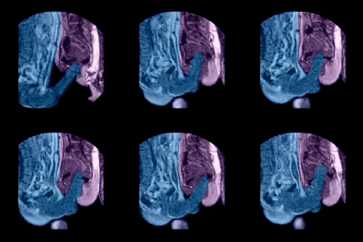
In 1999, a team of Dutch researchers led by gynaecologist Willibrord Schultz, University of Groningen, observed sexual intercourse with the aid of magnetic imaging. They discovered that the penis, thanks to the length of its root, adapts to the angle of the vagina. During copulation, the organ bends in the exact middle between tip and root so that it is parallel to the woman’s spine. Thanks to this flexibility, the tip of the penis can reach the uterine orifice, making the distance sperm cells have to travel to the egg cell as short as possible.
The study’s underlying concept was examined by the hospital’s ethics commission and approved with a few conditions. For example, the work had to be done separately from normal operations involving patients, and only staff would be permitted to participate which volunteered for it. Imaging began in 1991, but there was a major problem: 52 seconds of immobility were necessary! The Groningen hospital purchased a faster MRI scanner in 1996, but even the 12 seconds it required were still unfeasible. The availability of potency enhancer Viagra in 1998 finally made imaging possible. Thirteen attempts with eight couples finally produced successful results.
The images were published in the renowned British Medical Journal, and detailed reports appeared in Der Spiegel in addition to a number of other media. In 2000, Harvard University awarded the Dutch team the Ig Nobel Prize, which is given to research that “cannot or should not be reproduced”.
Sources:
Willibrord Weijmar Schultz, et al., Magnetic resonance imaging of male and female genitals during coitus and female sexual arousal, BMJ 1999, 319, 1596-1600
Jörg Blech, Leonardos falscher Pinsel [Leonardo’s fake brush], Der Spiegel, 3 Jan 2000, 148 f
Jörg Albrecht, Ruft beim Schuss ganz einfach Bumm [Just say boom when the shot’s been fired], Die Zeit – Wissen, 2000, 42

